Tag Archives: electron microscopy
Cs adsorption by Mn‒Fe-based Prussian blue analogs formed in agarose gel

The Fukushima nuclear accident triggered a massive release of radioactive cesium (Cs) isotopes into the environment and generated a large amount of contaminated water. Because Cs isotopes pose serious threats to the environment (particularly the aquatic environment),
Protein structure detection in intermediate-resolution cryo-EM maps using deep learning

Cryo-EM has established its position in structural biology as an indispensable method of choice for determining macromolecular structures due to recent technological breakthroughs. The recent years have observed a steep increase of biomolecular structures solved by cryo-EM,
Cryo-electron microscopy in drug development
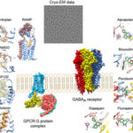
Cryo-electron microscopy (cryo-EM) is becoming the method of choice in structure determination of membrane proteins and has great potential for structure-based drug discovery (SBDD). Cryo-EM provides high-resolution structural information of a membrane protein without the need for
Dynamic electron microscopy: recording of ATP-induced myosin head movement in living muscle myosin filament

Muscle contraction results from relative sliding between actin and myosin filaments, caused by cyclic movement of myosin heads coupled with ATP hydrolysis. It is generally believed that individual myosin heads M), extending from myosin filaments, first bind
Some questions remain unanswered for alpha-Crystallin

α-Crystallin together with β- and γ-crystallins make about 40% of the dry weight of fibrous cells in eye and 90% of all proteins. α-Crystallin is responsible for the eye lens transparency creating the necessary refractive index (1.4
A “Cool” way to study Parkinson´s Disease

The Big Bang Theory left us amazed about how Leonard was able to bring back a snowflake embedded in resin from the North Pole to give to Penny. The reality is that this concept is not so
In vivo dissection of extracellular vesicles production in plants

In the last decade, the number of studies isolating and describing extracellular vesicles (EVs) has dramatically grown once their participation in intercellular communication has been confirmed. As transporters of bioactive molecules, EVs can take part in different
Limitations in nanoelectronics: current and temperature effects

Electrical cables are commonly used in our daily life. Typically they have metallic core covered with isolation. For example, one uses this wire to connect a lamp to a battery. When connected, electrons (negative elementary electric charges)
Data storage – Quo vadis?

The amount of digital data created by mankind is increasing exponentially. Big companies have evolved addressing the need to store and manage such data masses. Google, Microsoft, and others are challenged to save several petabytes (1 PB
Exploiting an ‘Achilles heel’ of DNA replication to arrest viral proliferation

In order to proliferate all organisms need to replicate their genomes. In most, this is achieved by a process named DNA replication by which a parental DNA gives rise to two faithful copies of itself. For this
A new type of mycovirus

Virus infects Aspergillus fumigatus, the fungus that can cause the human disease aspergillosis Researchers, led by Dr Robert Coutts, Leverhulme Research Fellow from the School of Life and Medical Sciences at the University of Hertfordshire, and Dr
A study to establishing the structural integrity of core−shell nanoparticles

Lanthanide – doped core – shell nanoparticles, such as NaYF4:Yb/Er@NaYF4, have attracted increasing attention for applications in photonics, photovoltaics, and biological imaging and therapeutics. When compared to conventional nanoparticles that suffer from extensive and uncontrollable dopant −


